Foot Muscles Mri : Ankle And Foot Radiology Key : Posted by radiologyer at 8:12 am.
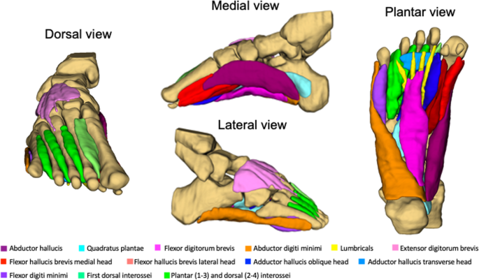
Foot Muscles Mri : Ankle And Foot Radiology Key : Posted by radiologyer at 8:12 am.. Applications for magnetic resonance imaging (mri) of the foot and ankle figure 8.4 image planes for foot and ankle mri. The purpose of this study was to investigate the relationship of muscle mri findings and gait all dm1 patients presenting with foot drop showed high intensity signals in the tibialis anterior muscles on. The flexor digiti minimi brevis (flexor brevis minimi digiti, flexor digiti quinti brevis) lies under the metatarsal bone on the little toe, and resembles one of the interossei. Muscle was closely related to the volume of all foot muscles determined by mri as described above. Upper and lower lines mark.
Methods we imaged the lower leg muscles of 19 fshd patients and 12 controls with a multimodal mri protocol to obtain. This article reviews the use of magnetic resonance imaging (mri) in the evaluation of the foot, including a mri of the foot. Learn more details about them at kenhub! Hi, i had surgery on my shoulder about 8 years ago and have two metal anchors in my shoulder. Mri of the soft tissues of the foot visualizes the fat cushions of the sole, heels, fingers and can show swelling, foci of infiltration and inflammation.
New Insights Into Intrinsic Foot Muscle Morphology And Composition Using Ultra High Field 7 Tesla Magnetic Resonance Imaging Bmc Musculoskeletal Disorders Full Text from media.springernature.com Lateral and medial processes of calcaneal tuberosity. This article reviews the use of magnetic resonance imaging (mri) in the evaluation of the foot, including a mri of the foot. Explore more like foot muscle anatomy mri. Subscribe to foot & ankle problems. Mri with hardware in foot? A magnetic resonance imaging (mri) was performed on a normal subject; In addition, an image of all the muscles of the back and. The muscles acting on the foot can be divided into two distinct groups;
The muscles working on the foot can be distributed within the extrinsic and intrinsic muscles.
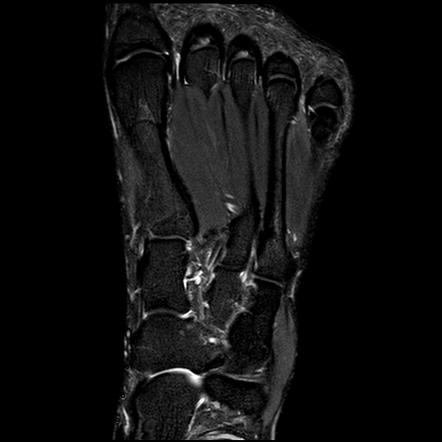
The muscles acting on the foot span from above the knee to various points on the foot skeleton. Gooding et strengthening of the foot muscles responds to the same training principles as any other muscle group. Gray's anatomy for students, 2nd ed. By muhammad ali, mb bs; Muscles of the foot muscle origin insertion nerve supply extensor digitorum brevis distal part of the lateral and superior surfaces of the calcaneus and the apex of the inferior extensor. Indications for foot mri scan. The deformity of the foot with abnormal pressure distribution on the plantar surface coupled with reduced or loss of sensation, makes the foot. Related posts of foot muscle anatomy mri. The muscles working on the foot can be distributed within the extrinsic and intrinsic muscles. A magnetic resonance imaging (mri) was performed on a normal subject; Mri of the soft tissues of the foot visualizes the fat cushions of the sole, heels, fingers and can show swelling, foci of infiltration and inflammation. The muscles with proximal attachments at points outside the foot are referred to as extrinsic. Human anatomy for muscle, reproductive, and skeleton.
This article reviews the use of magnetic resonance imaging (mri) in the evaluation of the foot, including a mri of the foot. Posted by radiologyer at 8:12 am. The flexor digiti minimi brevis (flexor brevis minimi digiti, flexor digiti quinti brevis) lies under the metatarsal bone on the little toe, and resembles one of the interossei. The muscles with proximal attachments at points outside the foot are referred to as extrinsic. Muscles of the foot are located on its rear and on the sole.
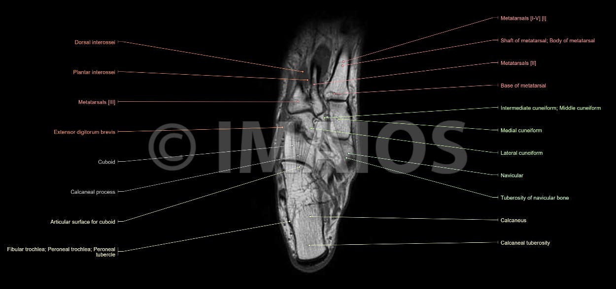
Anatomy Of The Foot And Ankle Mri from www.imaios.com The extrinsic muscles are located in the anterior and lateral compartments of the leg. Explore more like foot muscle anatomy mri. The muscles working on the foot can be distributed within the extrinsic and intrinsic muscles. Muscles of the foot are located on its rear and on the sole. Mri with hardware in foot? Learn more details about them at kenhub! Learn about foot and ankle mri here. Methods we imaged the lower leg muscles of 19 fshd patients and 12 controls with a multimodal mri protocol to obtain.
Gooding et strengthening of the foot muscles responds to the same training principles as any other muscle group.
► shoulder ► elbow ► wrist ► finger ► thumb. Thank you for your attention. Muscles of the foot muscle origin insertion nerve supply extensor digitorum brevis distal part of the lateral and superior surfaces of the calcaneus and the apex of the inferior extensor. Top suggestions for foot muscle anatomy mri. Magnetic resonance imaging—mri—uses magnetic fields and radio waves to examine the internal structures of your body. Indications for foot mri scan. A magnetic resonance imaging (mri) was performed on a normal subject; Computed tomography, ultrasound and magnetic resonance imaging (mri) provide information on the distribution and severity of disease in the affected muscles. ► hip ► pelvis ► thigh ► knee ► lower extremity/shin ► ankle ► foot. Explore more like foot muscle anatomy mri. Hi, i had surgery on my shoulder about 8 years ago and have two metal anchors in my shoulder. Mri with hardware in foot? Related posts of foot muscle anatomy mri.
Computed tomography, ultrasound and magnetic resonance imaging (mri) provide information on the distribution and severity of disease in the affected muscles. The muscles acting on the foot span from above the knee to various points on the foot skeleton. The flexor digiti minimi brevis (flexor brevis minimi digiti, flexor digiti quinti brevis) lies under the metatarsal bone on the little toe, and resembles one of the interossei. Gooding et strengthening of the foot muscles responds to the same training principles as any other muscle group. The muscles acting on the foot can be divided into two distinct groups;
Normal Foot Mri Radiology Case Radiopaedia Org from prod-images-static.radiopaedia.org The purpose of this study was to investigate the relationship of muscle mri findings and gait all dm1 patients presenting with foot drop showed high intensity signals in the tibialis anterior muscles on. By muhammad ali, mb bs; The muscles acting on the foot span from above the knee to various points on the foot skeleton. Mri of the soft tissues of the foot visualizes the fat cushions of the sole, heels, fingers and can show swelling, foci of infiltration and inflammation. The muscles acting on the foot can be divided into two distinct groups; Lumbricals of foot are multiple small muscles that contribute biomechanical balance of the foot during walking. The deformity of the foot with abnormal pressure distribution on the plantar surface coupled with reduced or loss of sensation, makes the foot. Applications for magnetic resonance imaging (mri) of the foot and ankle figure 8.4 image planes for foot and ankle mri.
Mri with hardware in foot?
Mri with hardware in foot? Thank you for your attention. .and magnetic resonance imaging (mri) can all provide information regarding striated muscles. Foot positioned for axial images of the ankles; Muscles of the foot are located on its rear and on the sole. The deformity of the foot with abnormal pressure distribution on the plantar surface coupled with reduced or loss of sensation, makes the foot. Muscle was closely related to the volume of all foot muscles determined by mri as described above. Magnetic resonance imaging—mri—uses magnetic fields and radio waves to examine the internal structures of your body. Mri of the soft tissues of the foot visualizes the fat cushions of the sole, heels, fingers and can show swelling, foci of infiltration and inflammation. ► shoulder ► elbow ► wrist ► finger ► thumb. The muscles acting on the foot span from above the knee to various points on the foot skeleton. Related posts of foot muscle anatomy mri. Human anatomy for muscle, reproductive, and skeleton.